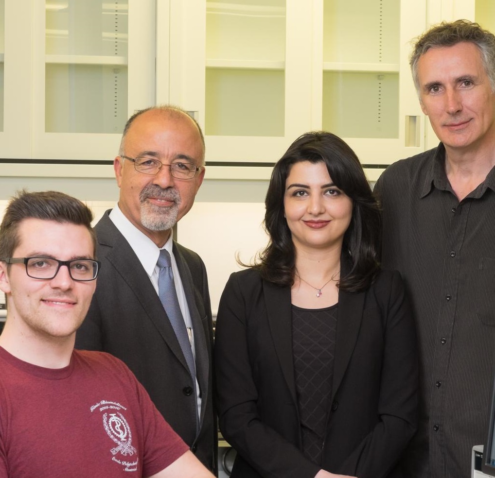
The Polystim Neurotechnologies lab at Polytechnique Montréal in collaboration with BioSA lab in the Lassonde School of Engineering at York University is helping Ghazal Nabovati develop an award-winning technology while equipping her with valuable multidisciplinary R&D skills.
The electrical engineering PhD candidate has created a “smart” petri dish that aims to redefine cell analysis. Her fully integrated cell imaging platform replaces traditional chemical-based assays with a reusable electronic biosensor that provides real-time imaging of growing cell cultures, and communicates information about their state of viability and proliferation.
The value of her experiential training was recognized last year, when she received the Brian L. Barge Award for Excellence in Microsystems Integration at the 2015 Innovation 360 TEXPO. Citing her work for its “significant and compelling economic potential with social benefits,” judges praised Nabovati for her use of multiple microsystems and disciplines, integrating biology, chip design, electronics, software and mechanical prototyping in a fully functioning imaging and analysis system.
The idea for the device grew out of her work with her supervisors, Dr. Mohamad Sawan, a Canada Research Chair in smart medical devices, director of the Polystim Lab, and a prolific innovator whose work integrates the disciplines of electrical engineering, psychology, neuroscience, biology and physiology; and Dr. Ebrahim Ghafar- Zadeh, Director of Biologically Inspired Sensors and Actuators (BioSA) Laboratory, and Assistant Professor in the Department of Electrical Engineering and Computer Science at York University.
“Current technologies are bulky, requiring a microscope and long sample preparation time,” Nabovati says. “They are labour-intensive and consume resources. My idea was to create a fully automated device that requires no labelling procedures; you just take the cells from the sample, put them on the device and right away you can see the results. There’s no need to wait.”
The core of the technology is a fully integrated CMOS capacitive sensor capable of detecting small charges around the cell membrane. The sensor converts this charge into a readable electronic signal, which is picked up and transmitted by the chip.
While simple and elegant in concept, the biosensor posed considerable challenges. Designing and making its individual parts was not nearly as difficult as making the parts work together, and creating a biocompatible package that is not toxic to cells, she says.
“It’s about the integration. That’s the tricky part,” Nabovati says. “It’s not just about making a chip that works. It’s how to match the chip with the biological environment—that’s the issue. It’s why there are no commercial devices in this space that are really efficient.”
It took her group six or seven months to solve the problem of protecting the cells from the chip and the chip from the cells. “That was the most challenging part.”
A key piece of the researchers’ work was proving that the biosensor worked. This meant finding a way to fabricate the biosensors and then validate them through experiments. “CMC helped me a lot,” Nabovati says. They provided her with access to Cadence and COMSOL software tools, and facilitated chip fabrication through TSMC of Taiwan. All of the fabrication process was done through CMC, and some of the equipment was made available through the Embedded Systems Canada (emSYSCAN) project, also managed by CMC.
She also credits her colleagues for their help. “Laurent Mouden provided important technical support, and Antoine Letourneau developed a real-time data acquisition system and helped with the biological experiments.”
There is still more work to be done on this technology; next steps include implementing microchambers on the chip, to provide different culture environments on the same chip.
“These micro-wells, fabricated on top of the sensors, allow us to perform high-throughput measurements such as cytotoxicity monitoring of anti-cancer drugs,” she says.
Lessons learned in developing the biosensor were broad in scope and extremely valuable, says Nabovati. “I gained a lot of experience, including designing microelectronic circuits. And I hadn’t had a chance to fab designs before, so it gave me the opportunity to learn about fabrication issues, layout, and implementation issues. The biology part was new for me as well—I learned a lot about cell culturing and cell manipulation.” Expecting to complete her PhD shortly, she hopes to take her expertise into industry.
Photo Credit: Yves Clement/Photo Features
July 2016

