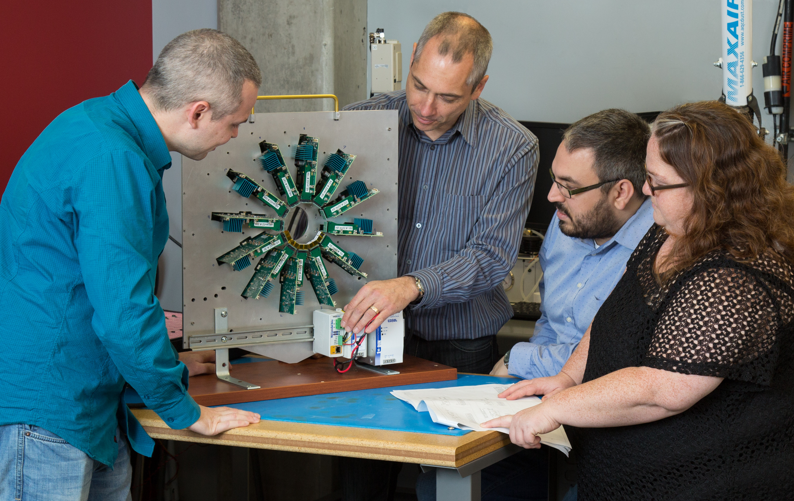
A powerful medical imaging system is becoming even more advanced thanks to the collaborative efforts of Université de Sherbrooke researchers Dr. Réjean Fontaine and Dr. Roger Lecomte.
The duo’s improvements to Positron Emission Tomography (PET) scanning achieved a breakthrough in 2012 by fully combining a PET and a computed tomography scanner using the same electronics. Their novel packaging and integration solution (“LabPET”) was honoured with the Canadian Manning Innovation Foundation’s 2012 Award of Distinction.
Further innovations have been reflected in their LabPET™ II, a second-generation compact scanner for small animals, from mice to rabbits, that sets a new standard for contrast-to-noise ratio and resolution in PET imaging. This enhanced performance was made possible because of a new generation of Application Specific Integrated Circuits (ASICs).
“For the last three years we have been working on a new ASIC with a 64-channel avalanche photodiode input,” says Fontaine, Professor of Electrical and Computer Engineering, and Director of the Research Group in Medical Devices (GRAMs). This feature is at the heart of LabPET II. He points out that the first detector device, introduced in 2006, collected 25 pixels per square centimetre with crystal scintillators that were about two millimetres wide. The new detector module collects 70 pixels per square centimetre with crystal scintillators that are about half as wide which enables a higher contrast in the image. This new detector required the collaboration of many other colleagues including professors J.F. Pratte, S. Charlebois, D. Danovitch from Université de Sherbrooke and A. Lahkssassi from Université du Québec en Outaouais.
Thanks to that high image contrast, information is generated much more quickly, animals spend less time being scanned, and areas of interest, such as the site of a tumour, can immediately be isolated. “However, we have a lot of electronics within a very small volume,” notes Fontaine, who says it was crucial to find a way of removing heat from the front-end electronics to keep
detectors at a stable temperature.
PET systems rely on injecting a radiotracer, a molecule labeled with a positron-emitting radioisotope, in a subject. The tracer targets specific chemical processes in cells or tissues where it is metabolized and the radiation emitted by the radioisotope is used to locate where the bioprocess occurred. The emitted positron (positively charged electron) gradually loses its kinetic energy as it travels in tissue and eventually interacts with a surrounding electron, generating two energetic annihilation photons that are emitted nearly back to back. This activity is captured by the detectors, which use the information to create a detailed image of the radioactivity distribution in the subject.
Unfortunately, because the positron is moving it is difficult to precisely determine the disintegration point of origin and this limits spatial resolution – a challenge in preclinical PET scanners.
“In practice, the exact radiotracer location may be off by as much as half a millimeter and up to three or four millimetres when you consider the non-collinearity effect in large human scanners,” says Fontaine. “That means you can hardly locate a cancerous tumour smaller than that. And if you want to see lesions in a subject as small as a rat or a mouse, you must have even higher spatial resolution than in clinical scanners.”
“In clinical PET, the main show-stopper is spatial resolution coming from the non-perfect collinearity of emitted annihilation photons and large scanner bore,” explains Fontaine. “However, a big breakthrough came about 10 years ago with time-of-flight technology.”
This innovation measures to a few hundreds of picoseconds the travel time for each annihilation photon from its point of origin to the detector. With this more precise information, higher contrast images can be reconstructed. Further development of this technology, by one order of magnitude, is still needed before it contributes to improvements in preclinical scanners, he says.
“The next breakthrough in preclinical PET scanners will be to include the time of flight information in the image reconstruction process, thanks to new detectors with higher performance,” says Fontaine. “It will make a huge difference to whole-body scanners because of what we’re developing for small animal imaging.”
The entire team credit their technological advances to CMC Microsystems, which provided the resarch group at Université de Sherbrooke with direct access to chip fabrication facilities that enabled the development of the powerful ASIC inside the LabPET II. CMC also enabled the team to build relationships with industry partners, who helped to meet the challenge of an extremely compact assembly.
To date, there have been two successful technology transfers of PET scanners, the LabPET I in 2006 and LabPET II in 2016. Lecomte is currently supporting the
creation of a new spinoff, iR&T Inc., which will market the LabPET II scanner to customers around the world.
These medical imaging breakthroughs can even be used as proof of concepts for broader application in “big science” projects, Fontaine says.
“The Large Hadron Collider at CERN, the Next Enriched Xenon Observatory (nEXO), or the Dark Matter Experiment using Argon Pulse-shape discrimination (DEAP) at SNOLAB, where my colleagues Charlebois and Pratte are actively involved, are examples of scientific spin-offs and research synergy,” says Fontaine.
Benefits are also accruing to the researchers’ graduate students, who are working on every aspect of PET projects. Recently, three of these students won international awards at the 2016 IEEE Nuclear Science Symposium and Medical Imaging Conference in Strasbourg, France, including the prestigious 2016 Radiation Instrumentation Early Career Achievement Award, for “significant and innovative technical contributions to the fields of radiation instrumentation and measurement techniques for ionizing radiation.” The award is given to a researcher less than 10 years after the completion of a PhD and Marc-André Tetrault was selected even before completing his PhD.
January 2017

